Our research focuses on developing novel acquisition, reconstruction, and data processing techniques across various MRI modalities, aimed at addressing critical challenges and advancing both clinical and neuroscientific applications.
Advanced MRI acquisition and encoding methods
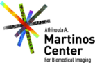
We develop advanced MRI acquisition techniques that leverage novel spatial encoding, data sampling, and contrast encoding methods with advanced reconstruction algorithms. One example is Echo Planar Time-resolved Imaging (EPTI), a next-gen EPI readout that produces distortion-free, T2/T2* blurring-free images while efficiently capturing rich multi-echo information. EPTI offers a new tool for diverse imaging applications, including fMRI and dMRI, and has been widely adopted by research institutions worldwide.
Functional MRI: ultra-high spatiotemporal resolution and precision functional mapping
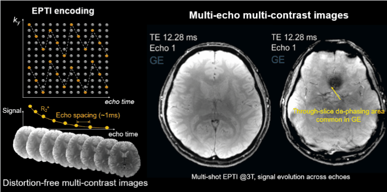
One theme of our fMRI research is achieving high-resolution fMRI aimed at improving neuronal specificity. For this, we have developed novel acquisition/reconstruction techniques that achieve significantly higher spatiotemporal resolutions (e.g., mesoscopic levels with <=0.1 µL voxels at high temporal resolution using TIDY) and obtain imaging contrasts that are more confined to microvascular signals. This enables investigation of human brain function at the level of cortical columns and layers—fundamental elements of brain organization and neural processing.
Another theme focuses on standard fMRI applications towards precision and individual functional mapping, where we have developed acquisitions that improve functional sensitivity and reliability by providing higher time-series fidelity and multi-echo physiological-denoising capabilities. A common aspect of our fMRI approaches in both themes also lies in addressing challenges that impede true spatiotemporal accuracy, including distortion, motion, and physiological noise. Our fMRI techniques are the core technologies for multiple NIH-funded projects and have been adopted by research institutions globally.
High-resolution diffusion MRI
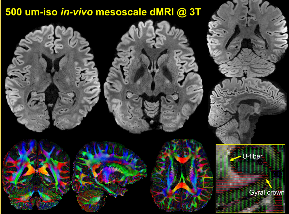
The in-vivo diffusion MRI (dMRI) acquisition and reconstruction technologies we are developing address major challenges, achieving improved SNR efficiency, motion robustness, and image quality, therefore pushing the limit of achievable resolution for in-vivo dMRI and enhancing its ability to probe tissue microstructures. For example, we have published the first-ever publicly-available in-vivo dMRI dataset sampled at submillimeter scales, which have been used by researchers worldwide to study vital brain circuitries relevant to Alzheimer’s disease, OCD, Parkinson’s disease, and more. Recently, our group has further developed in-vivo dMRI techniques that achieved mesoscopic resolutions at both 3T (500 µm iso) and 7T (485 µm iso) for the first time, as well as on the high-performance Connectome 2.0 scanner (387 µm iso), opening up exciting opportunities for studying exquisite in-vivo structures and connectivity.
4D CSF flow imaging for studying the glymphatic system

One focus of our research is the development of non-invasive imaging tools to map cerebrospinal fluid (CSF) flow in the human brain, aiming to gain new insights into the waste clearance system and glymphatic physiology. For instance, we have developed a novel CSF flowmetry technique with high sensitivity and specificity for mapping slow CSF flow. Using this technique, we demonstrated quantitative CSF flow mapping across the entire human brain, including the ventricles and the previously inaccessible yet critical subarachnoid space. Additionally, through the development of concurrent CSF flow and fMRI mapping technique, we have demonstrated the link between neural activities and CSF flow to better understand the role of neural processes in waste clearance.
Fast multi-parametric Quantitative MRI
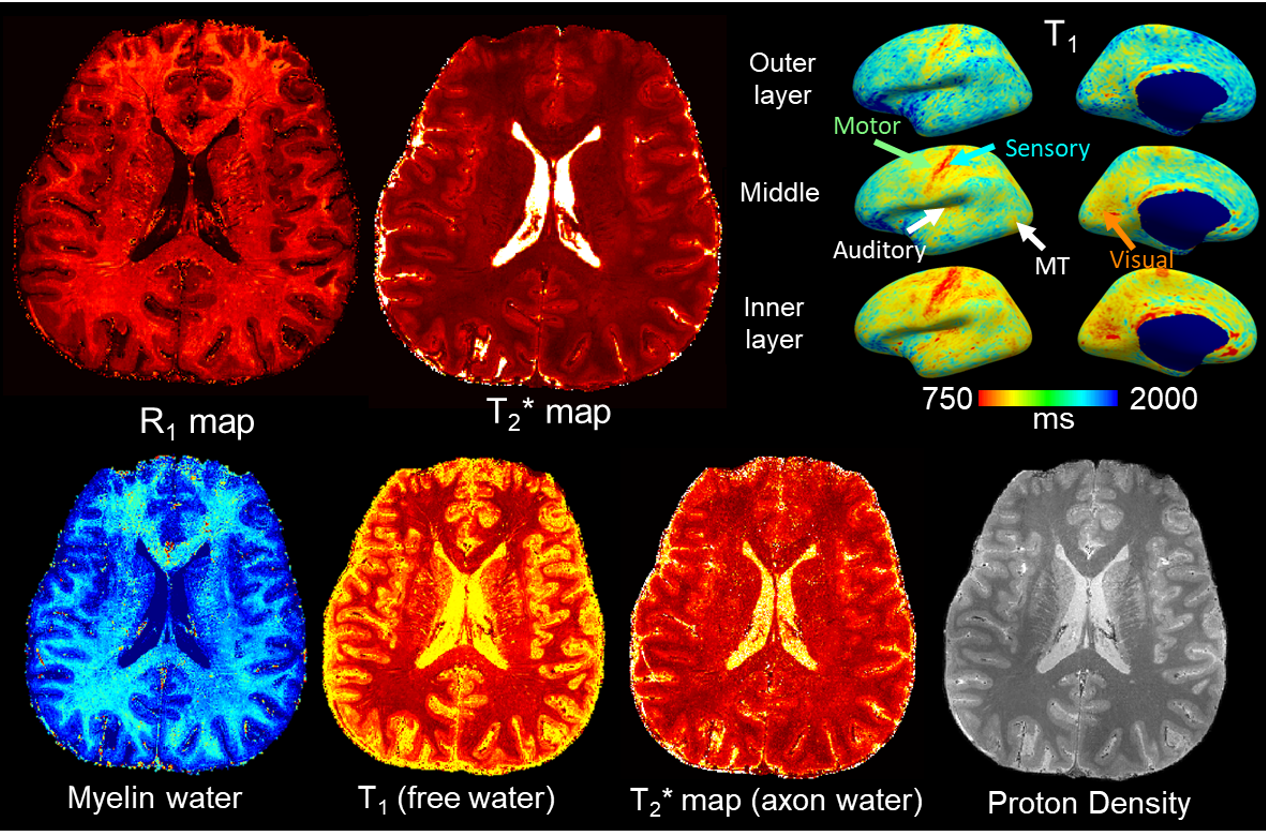
We are developing novel quantitative MRI techniques to address its major barriers toward clinical adoption and to probe tissue properties at fine time and spatial scales, including long scan time and motion vulnerability. For example, we designed an ultra-fast MR technique that enables whole-brain, multi-parametric quantitative MRI exams (T1, T2, T2*, PD) at 1-mm isotropic resolution in just 3 minutes with high repeatability and integrated motion-correction. In addition, our work on quantitative myelin water imaging achieves a fast 5-minute whole-brain scan and submillimeter imaging at 600 um iso in vivo, enabling detailed evaluation of cortical myeloarchitecture critical for studies of brain development and neurodegeneration studies.
Distortion-free multi-contrast Body Imaging
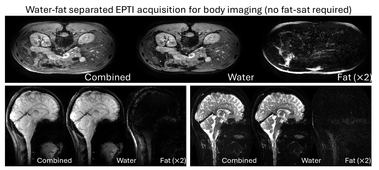
Building on the technologies developed for the brain, we are extending our novel acquisition and reconstruction strategies to body imaging, addressing challenges such as fat artifacts and severe image distortion and achieving high-resolution images. For example, by leveraging EPTI’s multi-echo readout for water-fat separation, we have successfully performed efficient multi-contrast and multi-parametric MRI at high spatial resolutions in various body regions, including the brain, neck, and abdomen.